


An ultrasound is a diagnostic procedure that is used to capture live images from the inside of your body. It is also referred to as sonography. Significantly, this test doesn’t use any radiation. This is why it is used mostly in women during pregnancy to view the developing baby. An ultrasound uses high-frequency sound waves to get pictures of tissues, organs, and other structures inside your body.
A sonographer performs this procedure. The images are further viewed by the radiologist or other specialists. Without making any incision or surgical cut in your skin, your doctor can see and diagnose multiple problems in your body.
Why it’s done
Mainly, ultrasonography is used for two purposes:
- To monitor the baby during pregnancy.
- To detect disorders in various body parts.
This test is used commonly to diagnose various conditions in the stomach and the reproductive parts. Moreover, an ultrasound is also very useful in monitoring the surgery (surgeons’ movements) during procedures like biopsy.
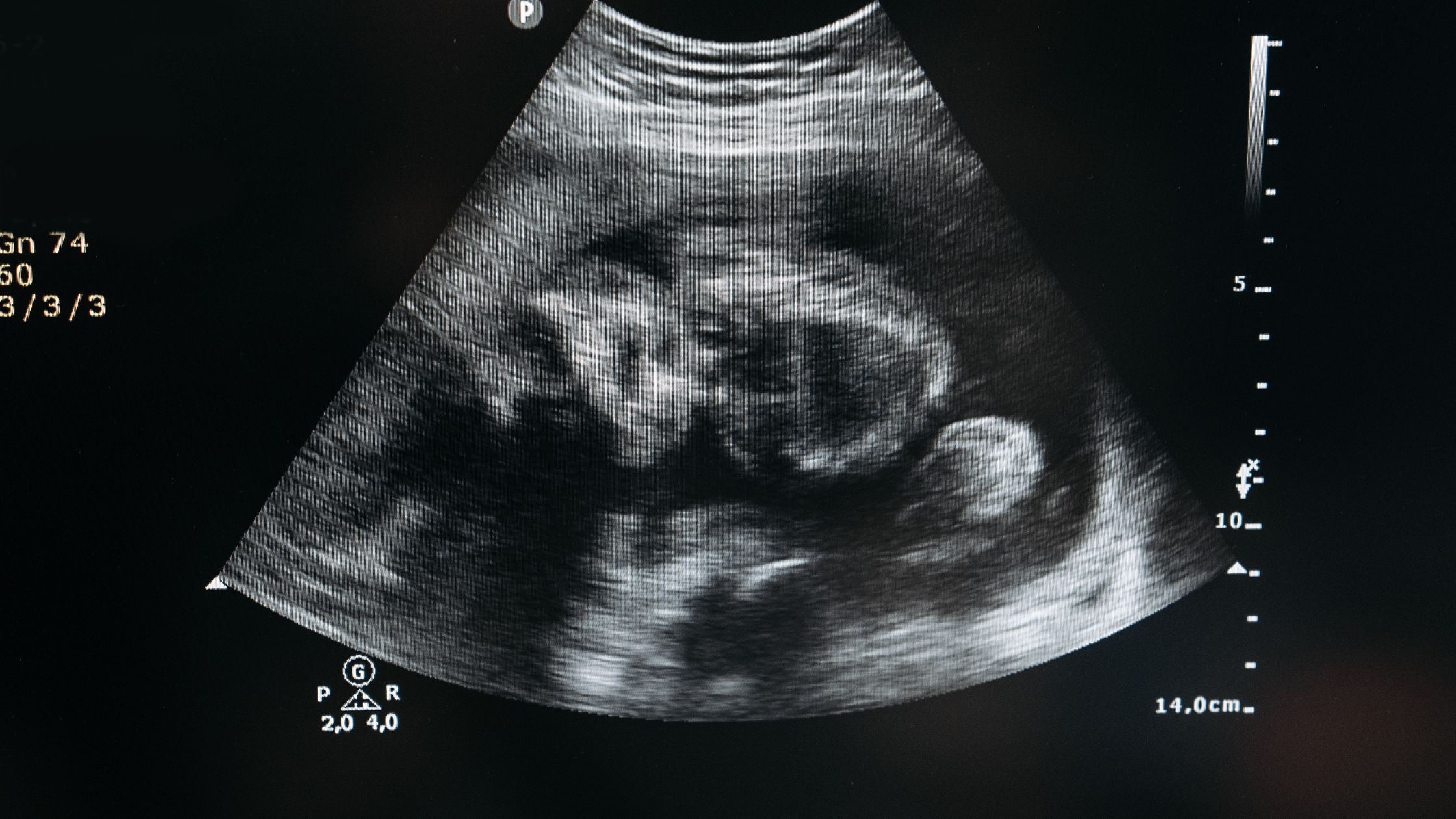
Pregnancy Ultrasound
A pregnancy ultrasound is used to view the growth of the developing baby and the reproductive organs of the mother. This test can screen for any problems during pregnancy. It helps the doctor in monitoring the development cycle of the fetus (unborn baby). Moreover, structural abnormalities of the fetus and blood flow can be monitored.
Your doctor may prescribe a pregnancy ultrasound:
- To confirm your pregnancy.
- To check the size of the fetus.
- To check the position of the fetus.
- To estimate your pregnancy duration.
- To investigate the signs of Down syndrome, in which the baby’s neck thickens.
- To investigate birth defects in different parts of the fetus’ body.
Here are the factors that are determined by a pregnancy ultrasound during different stages or trimesters of pregnancy.
- 6 to 11 weeks of pregnancy
- Confirmation of pregnancy
- Gestation of pregnancy, i.e., the start of the egg’s development
- The heartbeat of the embryo (fertilized egg)
- Determining if the pregnancy is safe or fatal
- 11 to 14 weeks of pregnancy
- Signs of Down’s syndrome in the baby (distinct appearance)
- Signs of heart disease in the baby (swelling and rapid heartbeat)
- 14 to 20 weeks of pregnancy
- Placenta’s position in the uterus
- Growth and position of the fetus
- Presence of amniotic fluid for baby’s growth
- 20 to 24 weeks of pregnancy
- Whether the baby is a single or twin
- The shape of the baby’s spinal cord and head
- The functioning of the baby’s heart and kidney
- 24 to 32 weeks of pregnancy
- The size and shape of the organs of the baby
- The position of the fetus
- The appearance of the fetus
- Blood flow to the fetus
Diagnostic Ultrasound
Your doctor may recommend an ultrasound if you experience pain in an organ for a long time. Diagnostic ultrasound is used to view different parts of the inside of your body and detect the problems associated with them. These may include the following.
- Testicles
- Uterus
- Ovaries
- Pancreas
- Eyes
- Gallbladder
- Kidneys
- Liver
- Spleen
- Thyroid
- Blood vessels
- Bladder
- Brain (in infants)
In women, diagnostic ultrasound is mainly used for:
- Diagnosing a breast lump that may be a cancer tumor.
- Investigating the cause of irregular periods or menstrual bleeding.
- Finding the cause of long-lasting pelvic pain.
- Diagnosing infertility.
In men, diagnostic ultrasound is mainly used for investigating the problems of the prostate gland and the reproductive tissues.
How it’s done
An ultrasound is an easy and safe diagnostic procedure that requires very little preparation. It is done by a sonography technician or sonographer.
Before the procedure
You may require specific preparations in accordance with the area of the body being tested.
- Especially, if your stomach is to be examined, your doctor may ask you to not eat anything for at least eight to ten hours before the procedure. This is because food (undigested) can interfere with the sound waves and lead to blur images.
- If your liver, pancreas, spleen, or gallbladder is to be examined, your doctor may ask you to not eat fat-containing food items the evening before the procedure and then fast till the procedure.
- If your pelvic area is to be examined, you may require a full bladder during the procedure. Your doctor will ask you to drink enough water before the ultrasound.
- You are allowed to drink water before the ultrasound and take the essential medications. However, make sure to tell your doctor about the prescribed drugs (including the cancer medicine, if any). This shall help in dose adjustments.
During the procedure
Before starting, you will be given a hospital gown to change into. The procedure may last for about 20 to 40 minutes.
- The sonographer shall use one of the Doppler flow machines. He may ask you to lie down on a table. The section of your body that is to be examined will be exposed for the procedure.
- Further, a special lubricating gel is applied to the area of the body to be examined. This jelly is used to prevent blur pictures. It can be easily removed after the test.
Once you are ready, the sonographer will press a transducer (small, microphone-like, and hand-held device) and move it to the area being examined. Majorly, a transducer works by sending sound waves. The waves it receives back are then sent to a computer to create live images.
- Depending upon the body part, the technician may ask you to change your positions during the test to get precise images.
After the procedure
- Once the procedure is done, the gel will be removed from your skin.
- You can resume back to your routine work after the ultrasound.
Results
The result of the ultrasound is given by a radiologist (imaging technician) who views the images. Such a picture is medically termed a sonogram. Your ultrasound report comprises the following:
- The technique used
- The impression
- The findings
- Comparison with previous test’s findings
Your radiologist will look at the pictures and check for:
- Fetus’ growth in the womb, if it was a pregnancy ultrasound.
- Swelling or inflammation, if it was a diagnostic ultrasound for investigating the cause of pain due to an accident.
- Visible growths or tumors (cancerous tissues), if it was a diagnostic ultrasound for cancer treatment.
- Functioning of the blood vessels, chambers, and other components of the heart, if it was a diagnostic ultrasound for heart problems.
- Abnormalities like decreased blood flow, swelling, breakage, and injuries, if it was a diagnostic ultrasound for any other part of the body.
Based on these observations and results, your doctor may recommend other tests like CT scan, MRI, and biopsy if the disease has not been precisely diagnosed.
Note: This article is not intended to be an alternative to medical advice, prescription, or appointment.